Respiratory A&P

Pin on Science class
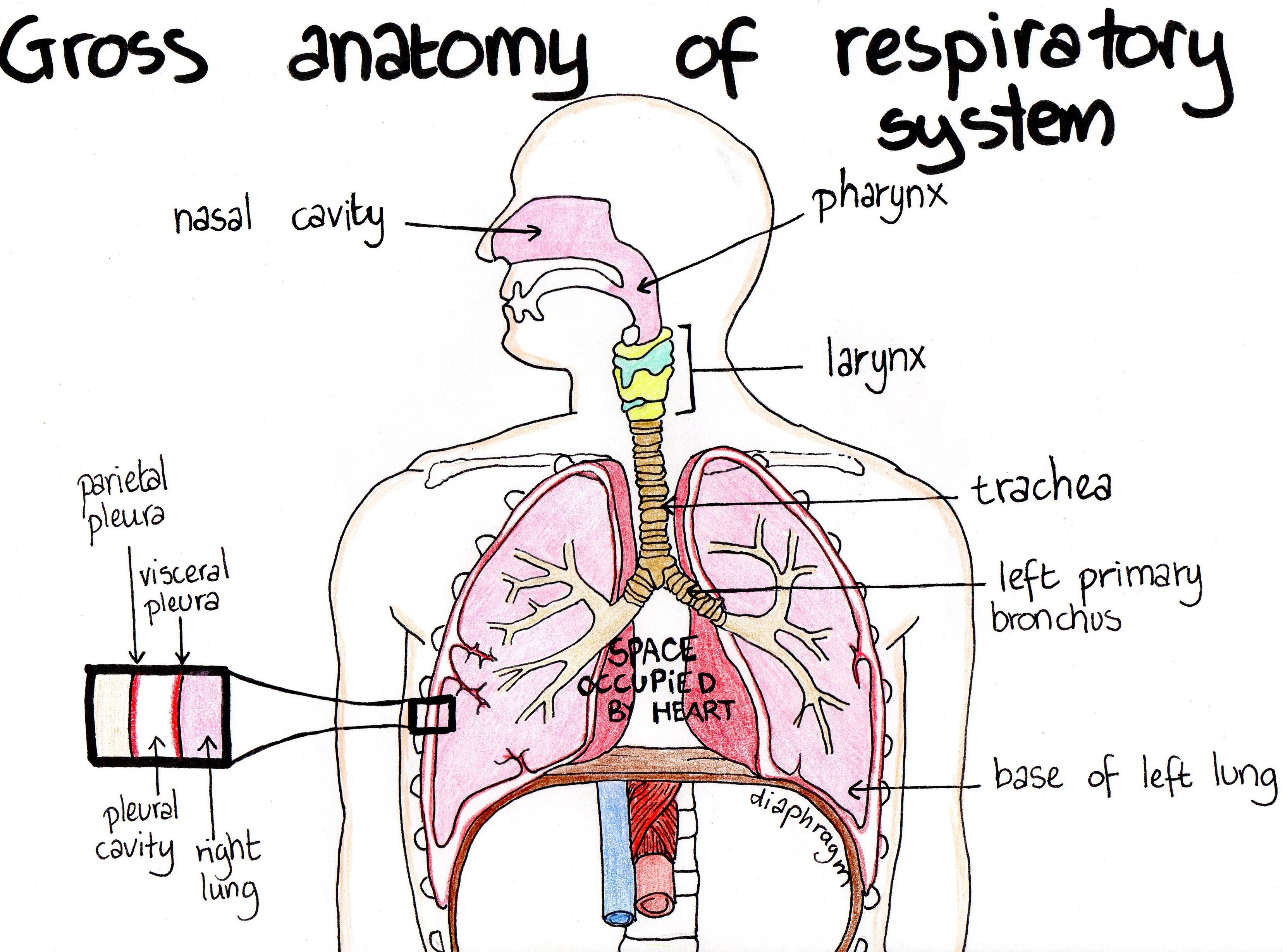
Diaphragm: The diaphragm is a broadband of muscle that sits underneath the lungs, attaching to the lower ribs, sternum, and lumbar spine and forming the base of the thoracic cavity. Respiratory system-related quizzes Respiratory System Anatomy Quiz (A Level) Mechanics of breathing GCSE Quiz Gaseous Exchange In The Lunges GCSE Quiz
The Upper Respiratory Tract and Mandibular Advancing Devices Dentist
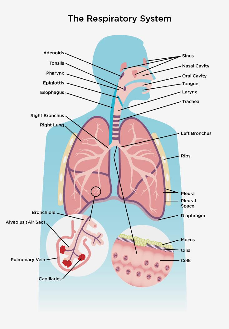
When cells use oxygen, they produce carbon dioxide and transfer it to your blood. Your bloodstream carries the carbon dioxide back to your lungs. When you exhale, you remove the carbon dioxide. Small hairs in your nose that act as an air-cleaning system and help filter out large particles. Mucus produced in your trachea and bronchial tubes to.

Labeled Diagram Of The Respiratory System For Kids Respiratory
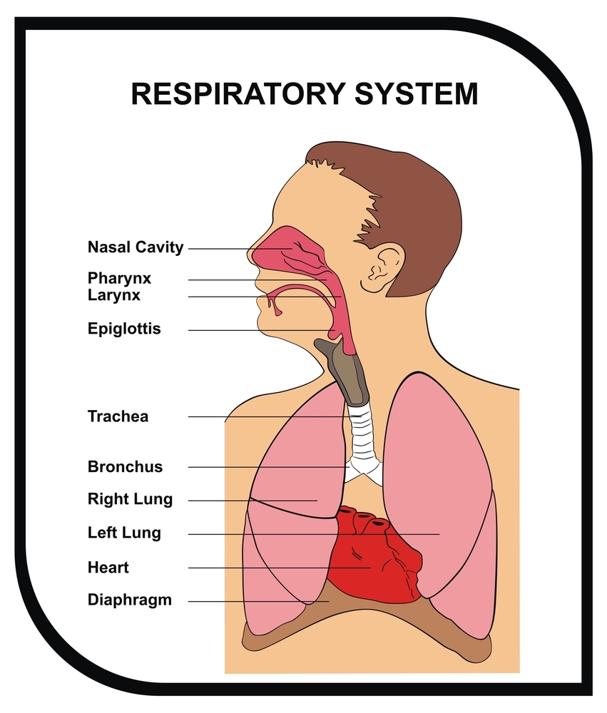
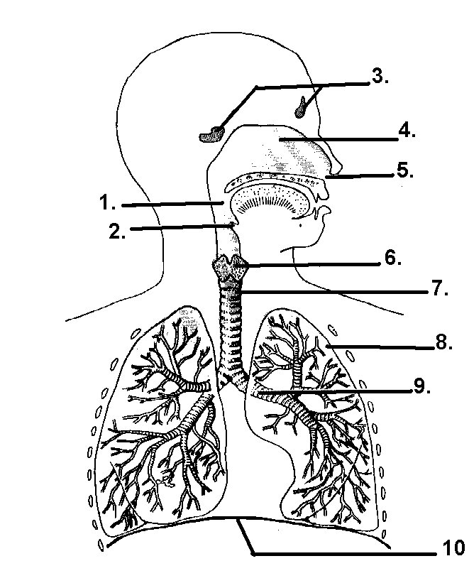
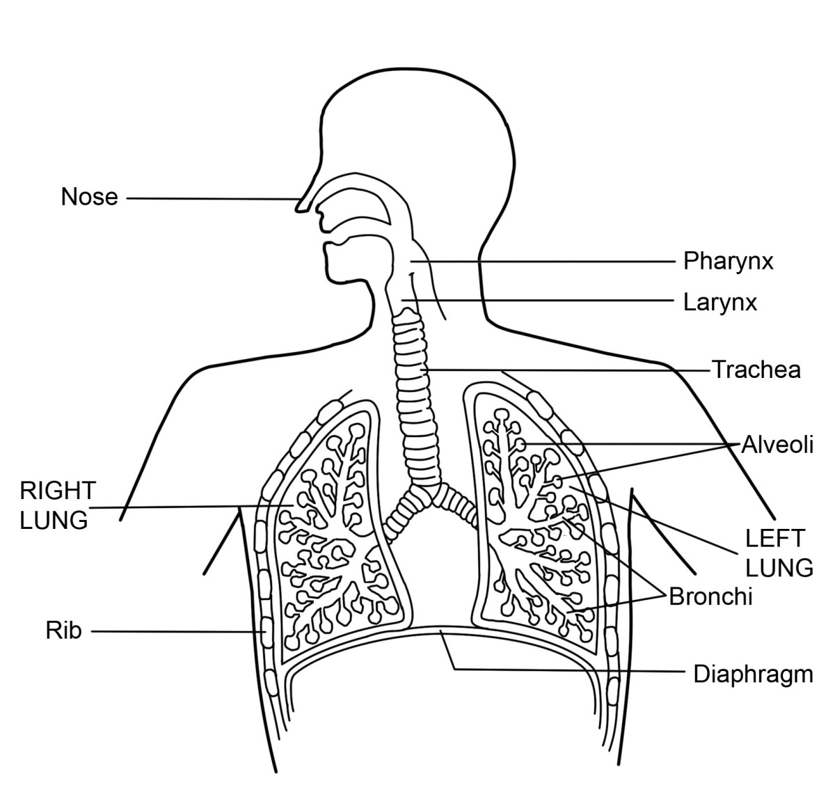
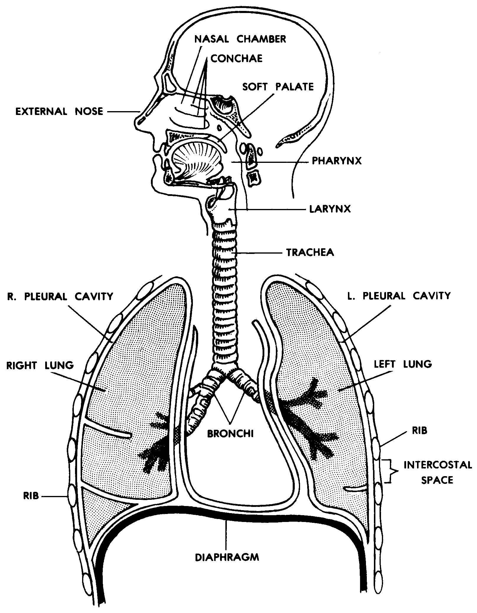
Diagram of the respiratory system. Air enters the body via the nose (preferably) or the mouth. The air enters the main windpipe, called the trachea, and continues en route to each lung via either the right or left bronchus (plural=bronchi). The lungs are separated into sections called lobes, two on the left and three on the right.
Respiratory System With Label Drawing at GetDrawings Free download
The major organs in the respiratory system are labeled. [Return to Figure 11.1]. Figure 11.2 image description: This figure shows a cross section view of the nose and throat. The major parts are labeled.. Diagram of the lungs with the major parts labeled (from top, clockwise): trachea, superior lobe, main bronchus, lobar bronchus,.

Respiratory System Diagrams To Label Human Respiratory System Diagram
All Osmosis Notes are clearly laid-out and contain striking images, tables, and diagrams to help visual learners understand complex topics quickly and efficiently. Find more information about Anatomy and Physiology of the Respiratory System: Respiratory system anatomy and physiology

Labeled Diagram Of The Respiratory System Of A Human
Respiratory system diagram Click on the interactive Bodymap below to move around the model and read more about the respiratory system. Parts of the respiratory system The respiratory.
Anatomy Of The Respiratory System Images Galleries
The lungs are the main part of your respiratory system. Here is how lungs work as the center of your breathing, the path a full breath takes in your body, and a 3-D model of lung anatomy.

Respiratory A&P
Your respiratory system is the network of organs and tissues that help you breathe. This system helps your body absorb oxygen from the air so your organs can work. It also cleans waste gases, such as carbon dioxide, from your blood. Common problems include allergies, diseases or infections.

C.1. Introduction to the Respiratory System
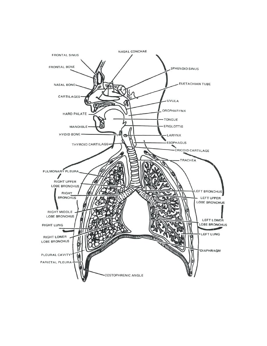
The upper respiratory tract refers to the parts of the respiratory system that lie outside the thorax, more specifically above the cricoid cartilage and vocal cords. It includes the nasal cavity, paranasal sinuses, pharynx and the superior portion of the larynx.
The Respiratory System Other Quiz Quizizz
human respiratory system, the system in humans that takes up oxygen and expels carbon dioxide. The design of the respiratory system Passage of air through the respiratory tract explained The respiratory tract conveys air from the mouth and nose to the lungs, where oxygen and carbon dioxide are exchanged between the alveoli and the capillaries.
Respiratory system HSC PDHPE
In the respiratory portion of the tract is connected to the bronchioles by alveolar ducts which in turn connect to alveoli. These are lined with simple squamosal epithelial. Unlike other epithelial tissue, the basement membrane of the tissue is connected to other squamosal epithelial cells; either other alveoli or capillaries. Figure 20.6. 20.6.
What is the Respiratory System Diagram and Function HubPages
Pelvic girdle and floor Female pelvis and reproductive organs Male pelvis and reproductive organs Urinary bladder and urethra Perineum Nerves, vessels and lymphatics of the pelvis Head and neck Overview Skull Face and scalp Infratemporal region and pterygopalatine fossa Orbit and contents Nasal region Ear Oral cavity Teeth Pharynx Neck Neuroanatomy
Respiratory System SJL Plymouth Tech Page
An ala is a cartilaginous structure that forms the lateral side of each naris (plural = nares), or nostril opening. The philtrum is the concave surface that connects the apex of the nose to the upper lip. Figure 22.3 Nose This illustration shows features of the external nose (top) and skeletal features of the nose (bottom).
Images 07. Respiratory System and Breathing Basic Human Anatomy
Lung anatomy can get quite complicated extremely quickly. Ease into the topic and cement your knowledge using Kenhub's respiratory system quizzes and labeled diagrams. Regulation of breathing. The breathing cycle is controlled by the respiratory centre located inside the medulla oblongata and the pons of the brain stem. Three major collections.
Respiratory System Diagram and Vocabulary MS. CRAWLEY
Inspiration (breathing in) The diaphragm contracts and moves downwards. The intercostal muscles contract and move the ribs upwards and outwards. This increases the size of the chest and decreases.

Schematic representation of the respiratory system. Download
Système Respiratoire Respiratory system This chart of the RESPIRATORY SYSTEM shows how you breathe. Breathing is the process that brings oxygen in the air into your lungs and moves oxygen and through your body.
